A cream-colored baroque-shaped shell blister with a light orange base, weighing 40.91 ct and measuring 22.79 × 20.59 × 13.21 mm, was recently submitted to GIA’s Mumbai laboratory for identification (figure 1). Externally, the large shell blister had a botryoidal arrangement with deep grooves. Viewed under 40× magnification, its high-luster surface displayed a typical nacreous structure of overlapping aragonite platelets. The base of the shell blister was sawn and polished, revealing concentric arcs and numerous polishing lines. Real-time X-ray microradiography (RTX) and X-ray computed microtomography (μ-CT) showed a distinct pattern of concentric growth arcs that terminated at the base. This was accompanied by an array of fine lines opposite the direction of the growth arcs forming grooves within the pearl (figure 2, A and B).
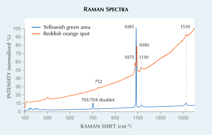
X-ray fluorescence (XRF) revealed a strong yellowish green reaction, indicative of its freshwater origin, with a small patch of reddish orange fluorescence observed at one end (figure 2C). Reddish orange fluorescence has been associated with vaterite in freshwater pearls (U. Wehrmeister et al., “Vaterite in freshwater cultured pearls from China and Japan,” Journal of Gemmology, Vol. 30, No. 7/8, 2007, pp. 399–412). Further chemical analysis using energy-dispersive X-ray fluorescence spectrometry was conducted on multiple spots. The yellowish green area yielded manganese levels ranging from 261 to 504 ppm and a strontium level of 413 to 452 ppm, while the reddish orange spot had a manganese level of 323 ppm and a strontium level of 415 ppm. Raman spectroscopy using 514 nm laser excitation on multiple surface spots revealed a combination of two calcium carbonate (CaCO3) polymorphs: aragonite and vaterite. The spot with reddish orange fluorescence had vaterite peaks at 752, 1075, and 1090 cm–1; the areas with a yellowish green fluorescence showed peaks at 701, 704, and 1085 cm–1, indicative of aragonite, along with polyenic pigment peaks at 1130 and 1530 cm–1 (figure 3). The photoluminescence spectra were consistent with the Raman and displayed strong fluorescence.
The presence of vaterite in association with aragonite in freshwater pearls has been extensively researched and recorded both in natural and cultured pearls. These minerals have also been occasionally documented in mollusk shells. It is therefore interesting to see such dual mineral phases in shell blisters as well. While it has often been speculated that vaterite serves as a precursor to aragonite during biomineralization, the presence of this mineral on the periphery suggests simultaneous formation within the shell blister (A.L. Soldati et al., “Structural characterization and chemical composition of aragonite and vaterite in freshwater cultured pearls,” Mineralogical Magazine, Vol. 72, No. 2, 2008, pp. 579–592). In certain cases, the presence of vaterite can be very difficult to detect through XRF analysis, especially when located within or around the core of the pearl. However, vaterite is more visible when the pearl is cut (Summer 2021 Gem News International, pp. 171–174).
Distinguishing between a natural blister pearl and a natural shell blister can be very challenging for laboratories due to the subtle variations in the definitions of these terms. Although such identifications are always debatable, the examined item was identified as a natural freshwater shell blister based mainly on its structural composition, external appearance, and distinct worked base, in addition to its notably large size.
A cream-colored baroque-shaped shell blister with a light orange base, weighing 40.91 ct and measuring 22.79 × 20.59 × 13.21 mm, was recently submitted to GIA’s Mumbai laboratory for identification (figure 1). Externally, the large shell blister had a botryoidal arrangement with deep grooves. Viewed under 40× magnification, its high-luster surface displayed a typical nacreous structure of overlapping aragonite platelets. The base of the shell blister was sawn and polished, revealing concentric arcs and numerous polishing lines. Real-time X-ray microradiography (RTX) and X-ray computed microtomography (μ-CT) showed a distinct pattern of concentric growth arcs that terminated at the base. This was accompanied by an array of fine lines opposite the direction of the growth arcs forming grooves within the pearl (figure 2, A and B).
X-ray fluorescence (XRF) revealed a strong yellowish green reaction, indicative of its freshwater origin, with a small patch of reddish orange fluorescence observed at one end (figure 2C). Reddish orange fluorescence has been associated with vaterite in freshwater pearls (U. Wehrmeister et al., “Vaterite in freshwater cultured pearls from China and Japan,” Journal of Gemmology, Vol. 30, No. 7/8, 2007, pp. 399–412). Further chemical analysis using energy-dispersive X-ray fluorescence spectrometry was conducted on multiple spots. The yellowish green area yielded manganese levels ranging from 261 to 504 ppm and a strontium level of 413 to 452 ppm, while the reddish orange spot had a manganese level of 323 ppm and a strontium level of 415 ppm. Raman spectroscopy using 514 nm laser excitation on multiple surface spots revealed a combination of two calcium carbonate (CaCO3) polymorphs: aragonite and vaterite. The spot with reddish orange fluorescence had vaterite peaks at 752, 1075, and 1090 cm–1; the areas with a yellowish green fluorescence showed peaks at 701, 704, and 1085 cm–1, indicative of aragonite, along with polyenic pigment peaks at 1130 and 1530 cm–1 (figure 3). The photoluminescence spectra were consistent with the Raman and displayed strong fluorescence.
The presence of vaterite in association with aragonite in freshwater pearls has been extensively researched and recorded both in natural and cultured pearls. These minerals have also been occasionally documented in mollusk shells. It is therefore interesting to see such dual mineral phases in shell blisters as well. While it has often been speculated that vaterite serves as a precursor to aragonite during biomineralization, the presence of this mineral on the periphery suggests simultaneous formation within the shell blister (A.L. Soldati et al., “Structural characterization and chemical composition of aragonite and vaterite in freshwater cultured pearls,” Mineralogical Magazine, Vol. 72, No. 2, 2008, pp. 579–592). In certain cases, the presence of vaterite can be very difficult to detect through XRF analysis, especially when located within or around the core of the pearl. However, vaterite is more visible when the pearl is cut (Summer 2021 Gem News International, pp. 171–174).
Distinguishing between a natural blister pearl and a natural shell blister can be very challenging for laboratories due to the subtle variations in the definitions of these terms. Although such identifications are always debatable, the examined item was identified as a natural freshwater shell blister based mainly on its structural composition, external appearance, and distinct worked base, in addition to its notably large size.